Valvular and flow phenotypes
Feature summary
LVOT aortic-root diameters
Aortic valve annulus, sinuses of Valsalva, and sinotubular junction diameters from LVOT cine segmentation.
Aortic insufficiency (I35.1)Marfan syndrome (Q87.4)Congenital aortic valve malformation (Q23.1)Disease badges are literature-context navigation only; not diagnoses, CardiacNexus classifiers, or validated phenotype-to-ICD associations.Mitral and tricuspid annular diameters
4-chamber segmentation-derived mitral and tricuspid annular minimum diameters at ED and ES, plus the ED TA/MA ratio.
Family: structuralUnit family: cm, unitlessSource: Cine long-axis CMRPrimary output group: ventricular_atrial_feature.csvAtrial fibrillation and flutter (I48)Nonrheumatic mitral valve disorders (I34)Nonrheumatic tricuspid valve disorders (I36)Disease badges are literature-context navigation only; not diagnoses, CardiacNexus classifiers, or validated phenotype-to-ICD associations.Aortic velocity, gradient, and flow volume
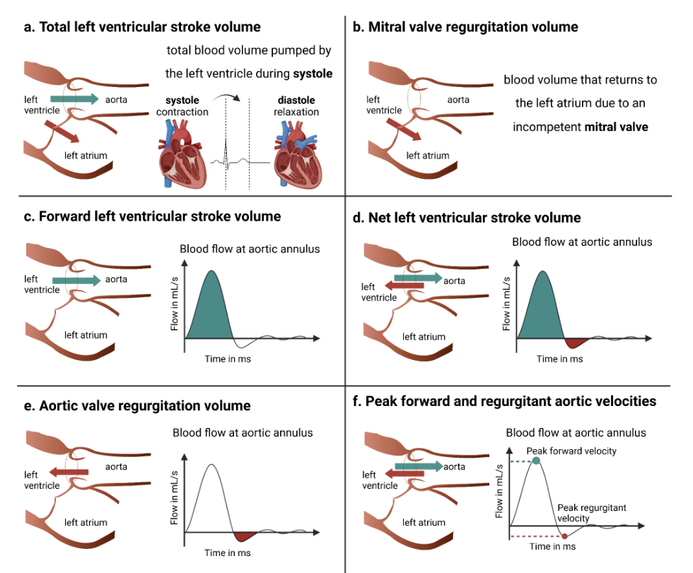
Phase-contrast aortic peak velocity, mean gradient, forward/backward flow, regurgitant fraction, and systolic reversal summaries.
Family: functionalUnit family: cm/s, mmHg, mL, "%"Source: Phase-contrast flow CMRPrimary output group: aortic_flow.csvAortic stenosis (I35.0)Aortic insufficiency (I35.1)Diastolic heart failure (I50.3)Disease badges are literature-context navigation only; not diagnoses, CardiacNexus classifiers, or validated phenotype-to-ICD associations.Aortic valve area, VTI, and flow displacement
Continuity-equation-style aortic valve area, velocity-time integral, and velocity-center displacement metrics from phase-contrast flow.
Family: functionalUnit family: cm², cm, "%"Source: Phase-contrast flow CMRPrimary output group: aortic_flow.csvAortic stenosis (I35.0)Aortic aneurysm and dissection (I71)Disease badges are literature-context navigation only; not diagnoses, CardiacNexus classifiers, or validated phenotype-to-ICD associations.
Valvular and flow phenotypes combine LVOT cine geometry, 4-chamber annular measurements, and phase-contrast aortic flow. This page is source-audited for the current CardiacNexus output names, but it treats valve disease labels as interpretation context only; no row is a stand-alone aortic stenosis, aortic regurgitation, mitral disease, or tricuspid disease diagnosis.
- Modality
- LVOT cine CMR, cine long-axis CMR, and phase-contrast flow CMR
- UKB source
- Data Fields 20212, 20208, and 20213
- Pipeline step
- LVOT aortic-root diameter extraction, combined valve landmark extraction, phase-contrast velocity and flow analysis
- Outputs
- LVOT.csv, ventricular_atrial_feature.csv, aortic_flow.csv, timeseries/aorta.npz, LVOT and flow QC plots
- Maturity
- Source-audited phenotype page
Clinical question
This page answers three separate questions: how large the aortic root is, how large the mitral and tricuspid annular spans are in 4-chamber cine, and how blood crosses the aortic phase-contrast plane. These are related to valve disease, aortopathy, and flow remodeling, but their acquisition planes and formulas differ enough that they should not be collapsed into one valve label.
Anatomical and physiological definition
The LVOT cine route measures aortic valve annulus, sinuses of Valsalva, and sinotubular junction diameters. The combined long-axis route measures mitral and tricuspid annular minimum diameters on 4-chamber segmentation at ventricular ED and ES.
Phase-contrast flow converts phase images into through-plane velocity, integrates velocity over the segmented aortic lumen, and summarizes area, velocity, gradient, forward/backward flow, valve area, VTI, systolic flow reversal, and flow displacement. Mean gradient is derived from velocity with a simplified Bernoulli relationship, while aortic valve area uses forward flow divided by VTI [2] [3].
Source acquisition and UKB field
LVOT cine measurements use UK Biobank Data Field 20212. Mitral and tricuspid annular measurements use the long-axis 4-chamber segmentation from Data Field 20208 plus ventricular timing from the short-axis timeseries. Aortic phase-contrast flow uses Data Field 20213 and relies on VENC metadata from the DICOM CSA header.
What exactly CardiacNexus measures
LVOT aortic-root diameters
eval_LVOT.py segments the LVOT cine frame stack and computes distances between landmarks for the aortic valve annulus, aortic sinuses, and sinotubular junction. The emitted rows are median diameters across accepted frames, with BSA-indexed versions when BSA is available.
Mitral and tricuspid annular diameters
eval_ventricular_atrial_feature.py calls evaluate_valve_length on the 4-chamber segmentation at ED and ES. It writes tricuspid and mitral annular minimum diameters at ED and ES, and writes the ED TA/MA ratio only when the ratio is not implausibly high.
Phase-contrast aortic flow
eval_phase_contrast.py computes aortic area from the phase-contrast segmentation, converts phase to velocity using VENC, integrates flow, estimates peak systole and end systole from the flow curve, and writes velocity, gradient, flow-volume, VTI, valve-area, flow-displacement, and systolic reversal summaries.

Aortic valve area, VTI, and flow displacement
The current aortic valve area row is derived from forward flow divided by VTI. Flow displacement compares the velocity-weighted flow center with the lumen center and normalizes by radius. These rows are method-sensitive and should be interpreted with plane prescription, segmentation, and velocity-aliasing context.
Output columns and units
CardiacNexus writes this page's current output families into LVOT.csv, ventricular_atrial_feature.csv, and aortic_flow.csv.
LVOT and annular structure
| Display family | Exact output column | Unit | Status | Schema note |
|---|---|---|---|---|
| LVOT diameter | LVOT: Aortic Valve Annulus Diameter [mm] | mm | current | median accepted-frame distance |
| LVOT diameter | LVOT: Aortic Sinuses Diameter [mm] | mm | current | median accepted-frame distance |
| LVOT diameter | LVOT: Sinotubular Junction Diameter [mm] | mm | current | median accepted-frame distance |
| LVOT indexed diameter | LVOT: Aortic Valve Annulus Diameter/BSA [mm/m^2] | mm/m^2 | conditional current output | requires BSA |
| LVOT indexed diameter | LVOT: Aortic Sinuses Diameter/BSA [mm/m^2] | mm/m^2 | conditional current output | requires BSA |
| LVOT indexed diameter | LVOT: Sinotubular Junction Diameter/BSA [mm/m^2] | mm/m^2 | conditional current output | requires BSA |
| Annular diameter | Valve: Tricuspid_diameter_ED [cm] | cm | current | 4-chamber ED segmentation |
| Annular diameter | Valve: Mitral_diameter_ED [cm] | cm | current | 4-chamber ED segmentation |
| Annular ratio | Valve: TA/MA Ratio_ED | unitless | conditional current output | skipped if implausibly high |
| Annular diameter | Valve: Tricuspid_diameter_ES [cm] | cm | current | 4-chamber ES segmentation |
| Annular diameter | Valve: Mitral_diameter_ES [cm] | cm | current | 4-chamber ES segmentation |
Phase-contrast aortic flow
| Display family | Exact output column | Unit | Status | Schema note |
|---|---|---|---|---|
| Aortic area | Aortic Flow: Maximum Area [mm^2] | mm^2 | current | maximum segmented lumen area |
| Aortic area | Aortic Flow: Minimum Area [mm^2] | mm^2 | current | minimum segmented lumen area |
| Aortic timing | Aortic Flow: T_ES [frame] | frame | current | end-systolic frame from flow curve |
| Aortic timing | Aortic Flow: T_peak_systole [frame] | frame | current | peak-systolic frame |
| Aortic timing | Aortic Flow: T_ES [ms] | ms | current | end-systolic time |
| Aortic timing | Aortic Flow: T_peak_systole [ms] | ms | current | peak-systolic time |
| Aortic velocity | Aortic Flow: Peak Velocity [cm/s] | cm/s | current | maximum velocity before ES |
| Aortic gradient | Aortic Flow: Mean Gradient [mmHg] | mmHg | current | simplified Bernoulli over systolic frames |
| Aortic flow | Aortic Flow: Forward Flow [mL] | mL | current | positive integrated flow |
| Aortic flow | Aortic Flow: Backward Flow [mL] | mL | current | reverse integrated flow |
| Aortic flow | Aortic Flow: Regurgitant Fraction [%] | % | current | backward/forward flow; >100% skipped |
| Aortic valve area | Aortic Flow: Aortic Valve Area [cm^2] | cm^2 | conditional current output | skipped when VTI exceeds QC threshold |
| Aortic VTI | Aortic Flow: Velocity Time Integral [cm] | cm | conditional current output | written when VTI passes QC threshold |
| Flow displacement | Aortic Flow: Flow Displacement Systolic Average [%] | % | current | systolic velocity-center displacement |
| Flow displacement | Aortic Flow: Flow Displacement Late Systolic Average [%] | % | current | late-systolic displacement |
| Flow displacement | Aortic Flow: Flow Displacement Diastolic Average [%] | % | current | diastolic displacement |
| Systolic reversal | Aortic Flow: Systolic Forward Flow [mL] | mL | current | systolic positive flow |
| Systolic reversal | Aortic Flow: Systolic Reverse Flow [mL] | mL | current | systolic reverse flow |
| Systolic reversal | Aortic Flow: Systolic Flow Reversal Ratio [%] | % | conditional current output | skipped when >50% |
Output reconciliation
| Evidence layer | Result |
|---|---|
| Implementation source | LVOT, phase-contrast flow, and 4-chamber annular rows checked against eval_LVOT.py, eval_phase_contrast.py, and eval_ventricular_atrial_feature.py |
| Output inventory | docs/data/output_column_inventory.yml records LVOT, aortic_flow, and ventricular_atrial_feature artifact families |
| Phenotype dictionary | docs/data/phenotype_dictionary.yml links promoted valvular/flow rows to this page |
| Page output table | promoted dictionary-backed rows and broader phase-contrast output families are listed above |
Required upstream inputs
- LVOT cine image and segmentation for aortic-root diameters;
- long-axis 4-chamber image and segmentation for annular diameters;
- ventricular and atrial aggregate CSVs, plus
timeseries/ventricle.npzandtimeseries/atrium.npz, for combined 4-chamber outputs; - phase-contrast magnitude/phase images, aortic segmentation, and DICOM VENC metadata for aortic flow;
- BSA table only for indexed LVOT diameter outputs.
Reference ranges with cohort and method context
| Feature | Source | Cohort | Reference value | Status | Note |
|---|---|---|---|---|---|
| Aortic root diameters | CMR aortic-root reference context [1] | CMR reference cohorts | method-dependent | Verified context source | Useful context, but CardiacNexus uses automated LVOT segmentation and median accepted-frame distances |
| Phase-contrast flow method | Phase-contrast CMR review [2] | method review | not a normal range | Verified context source | Used for velocity/flow method context |
| Valvular function at UKB scale | Kany et al. [3] | UKB-scale flow cohort | source-specific values | Verified context source | Do not display as direct thresholds without table-row extraction and method adjudication |
| Dynamic aortic flow volumes | Gomes et al. [4] | UKB-scale flow/genetics cohort | source-specific values | Verified context source | Regurgitant-fraction context, not a diagnostic cutoff in this page |
| Flow displacement | Sigovan et al. and Zhao et al. [5] [6] | 4D-flow / aortic-flow cohorts | method-dependent | Verified context source | Current CardiacNexus row is 2D phase-contrast-derived and needs method labels |
| Valve guideline thresholds | AHA/ACC valve guideline context [7] | clinical guideline | severe AS/AR thresholds are clinical-context dependent | Guideline context | Page cites only as clinical context, not as an automated classifier |
Source-located registry status: reference_range_sources.yml maps aortic-root and valve-flow context to CMR root, phase-contrast, UKB flow, regurgitant fraction, and flow-displacement sources. Guideline thresholds are not promoted as CardiacNexus thresholds because plane placement, VENC, phase-offset behavior, valve morphology, and clinical report context are outside the automated output contract.
Disease interpretation
Aortic-root dilation can contribute to aortic regurgitation and aortopathy interpretation, including bicuspid-valve and connective-tissue disease contexts. Peak velocity, mean gradient, VTI, and valve area are clinically relevant to aortic stenosis, while forward/backward flow and regurgitant fraction are relevant to aortic regurgitation [3] [7].
Mitral and tricuspid annular rows describe 4-chamber geometry and are useful in remodeling context, especially when read with atrial and ventricular size. Flow displacement and systolic reversal rows are eccentric-flow and hemodynamic context signals, not stand-alone disease labels [5] [6].
QC caveats and maturity boundary
LVOT diameter outputs depend on aortic-root segmentation quality and accepted-frame selection. Annular diameters depend on 4-chamber segmentation, ED/ES timing, and landmark geometry. Phase-contrast outputs depend on VENC, plane prescription, phase-to-velocity conversion, segmentation of the aortic lumen, aliasing, background phase behavior, and reliable flow-curve timing.
The current implementation skips or warns on implausible regurgitant fraction, high VTI, very high systolic flow reversal ratio, and unstable annular ratios. These are cohort phenotyping outputs and require review of QC plots and acquisition context before clinical interpretation.
Implementation provenance
| Feature family | Formula or computational route | Exact output columns | Source code file and function | Upstream dependencies | Conditional behavior | QC artifacts | Schema debt |
|---|---|---|---|---|---|---|---|
| LVOT aortic-root diameters | LVOT segmentation landmarks, median accepted-frame distances, optional BSA normalization | LVOT:* Diameter [mm], LVOT:* Diameter/BSA [mm/m^2] | eval_LVOT.py | lvot.nii.gz, seg_lvot.nii.gz, BSA table for indexed rows | indexed rows require BSA; frames can fail LVOT QC | visualization/aorta/LVOT_*.png | artifact key is uppercase LVOT in the aggregate registry |
| Annular diameters | 4-chamber ED/ES valve-length helper on segmentation-derived landmarks | Valve: Tricuspid_diameter_ED [cm], Valve: Mitral_diameter_ED [cm], Valve: TA/MA Ratio_ED, Valve: Tricuspid_diameter_ES [cm], Valve: Mitral_diameter_ES [cm] | eval_ventricular_atrial_feature.py; evaluate_valve_length | la_4ch.nii.gz, seg4_la_4ch.nii.gz, ventricle.npz, ventricular and atrial aggregate CSVs | TA/MA ratio skipped when implausibly high; subject skipped if upstream aggregates/timeseries missing | visualization/combined/valve_ED.png, visualization/combined/valve_ES.png | combined artifact also emits AV/IPVT rows that belong mainly to cross-chamber interpretation |
| Aortic velocity and flow | phase image to velocity using VENC, lumen integration, peak/ES timing from flow curve | velocity, gradient, forward/backward flow, regurgitant fraction, timing rows | eval_phase_contrast.py | phase-contrast images, segmentation, DICOM CSA VENC metadata | subject skipped if VENC/inputs fail; regurgitant fraction >100% skipped | phase overlays, velocity and flow time-series plots, timeseries/aorta.npz | plane and velocity correction context is not encoded in the output labels |
| Aortic valve area and VTI | systolic VTI from peak-velocity curve; valve area from forward flow divided by VTI | Aortic Flow: Aortic Valve Area [cm^2], Aortic Flow: Velocity Time Integral [cm] | eval_phase_contrast.py | same phase-contrast flow inputs | VTI rows skipped if VTI exceeds QC threshold | velocity/VTI plots | continuity-equation interpretation requires method context |
| Flow displacement and reversal | velocity-center displacement normalized by lumen radius; systolic forward/reverse flow summaries | Aortic Flow: Flow Displacement*, Aortic Flow: Systolic* rows | eval_phase_contrast.py | segmented lumen and phase-derived velocity field | systolic reversal ratio skipped when >50% | flow-displacement and rotation-angle plots | displacement rows are sensitive to noisy contours and 2D plane choice |
Source audit
- Current LVOT output labels and BSA conditionals were checked against
eval_LVOT.py. - Current phase-contrast area, timing, velocity, gradient, flow, valve-area, VTI, flow-displacement, and systolic reversal labels were checked against
eval_phase_contrast.py. - Current annular diameter and TA/MA ratio labels were checked against
eval_ventricular_atrial_feature.py. docs/data/reference_sources.ymlis present and used as the curated reference-source registry for this page.- Textbook context boundary: page-specific code, method literature, and guideline context are sufficient for draft rollout; Braunwald/Hurst background was not needed for the phenotype-specific output contract.
- One regurgitant-fraction schematic is displayed from a page-local public path and registered in
docs/data/figure_provenance.yml; permission and exact source-panel review remain pending for draft use.
Related pages
- Aortic structure
- Aortic stiffness and distensibility
- Cross-chamber phenotypes
- LVOT cine imaging
- Phase-contrast flow imaging
References
- Kawel-Boehm N, Hetzel SJ, Ambale-Venkatesh B, Captur G, Francois CJ, Jerosch-Herold M, Salerno M, Teague SD, Valsangiacomo-Buechel ER, Van Der Geest RJ, Bluemke DA. Reference ranges for cardiovascular magnetic resonance in adults and children: 2020 update. Journal of Cardiovascular Magnetic Resonance. 2020;22(1):87.
- Nayak KS, Nielsen JF, Bernstein MA, Markl M, Gatehouse PD, Botnar RM, Saloner D, Lorenz C, Wen H, Hu BS, Epstein FH, Oshinski JN, Raman SV. Cardiovascular magnetic resonance phase contrast imaging. Journal of Cardiovascular Magnetic Resonance. 2015;17(1):71.
- Kany S, Ramo JT, Hou C, Jurgens SJ, Nauffal V, Cunningham J, Lau ES, Butte AJ, Ho JE, Olgin JE, Elmariah S, Lindsay ME, Ellinor PT, Pirruccello JP. Assessment of valvular function in over 47,000 people using deep learning-based flow measurements. Cardiovascular Medicine preprint. 2023.
- Gomes B, Singh A, O'Sullivan JW, Amar D, Kostur M, Haddad F, Salerno M, Parikh VN, Meder B, Ashley EA. Genetic architecture of cardiac dynamic flow volumes. Cardiovascular Medicine preprint. 2022. doi:10.1101/2022.10.05.22280733.
- Sigovan M, Hope MD, Dyverfeldt P, Saloner D. Comparison of four-dimensional flow parameters for quantification of flow eccentricity in the ascending aorta. Journal of Magnetic Resonance Imaging. 2011;34(5):1226-1230.
- Zhao X, Garg P, Assadi H, Tan RS, Chai P, Yeo TJ, Matthews G, Mehmood Z, Leng S, Bryant JA, Teo LLS, Ong CC, Yip JW, Tan JL, van der Geest RJ, Zhong L. Aortic flow is associated with aging and exercise capacity. European Heart Journal Open. 2023;3(4):oead079. doi:10.1093/ehjopen/oead079. PMID:37635784; PMCID:PMC10460199.
- Nishimura RA, Otto CM, Bonow RO, Carabello BA, Erwin JP, Guyton RA, O'Gara PT, Ruiz CE, Skubas NJ, Sorajja P, Sundt TM, Thomas JD. 2014 AHA/ACC guideline for the management of patients with valvular heart disease. Journal of the American College of Cardiology. 2014;63(22):e57-e185.